Anatomy of the mammalian eye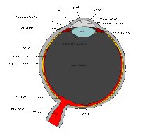 Schematic diagram of the human eyeThree layers
The structure of the mammalian eye can be divided into three main layers or tunics whose names reflect their basic functions: the fibrous tunic, the vascular tunic, and the nervous tunic.
- The fibrous tunic, also known as the tunica fibrosa oculi, is the outer layer of the eyeball consisting of the cornea and sclera.The sclera gives the eye most of its white color. It consists of dense connective tissue filled with the protein collagen to both protect the inner components of the eye and maintain its shape.
- The vascular tunic, also known as the tunica vasculosa oculi, is the middle vascularized layer which includes the iris, ciliary body, and choroid.The choroid contains blood vessels that supply the retinal cells with necessary oxygen and remove the waste products of respiration. The choroid gives the inner eye a dark color, which prevents disruptive reflections within the eye.
- The nervous tunic, also known as the tunica nervosa oculi, is the inner sensory which includes the retina. The retina contains the photosensitive rod and cone cells and associated neurons. To maximise vision and light absorption, the retina is a relatively smooth (but curved) layer. It does have two points at which it is different; the fovea and optic disc. The fovea is a dip in the retina directly opposite the lens, which is densely packed with cone cells. It is largely responsible for color vision in humans, and enables high acuity, such as is necessary in reading. The optic disc, sometimes referred to as the anatomical blind spot, is a point on the retina where the optic nerve pierces the retina to connect to the nerve cells on its inside. No photosensitive cells whatsoever exist at this point, it is thus "blind".
Anterior and posterior segments
The mammalian eye can also be divided into two main segments: the anterior segment and the posterior segment.
Anterior segment
The anterior segment is the front third of the eye that includes the structures in front of the vitreous humour: the cornea, iris, ciliary body, and lens. Within the anterior segment are two fluid-filled spaces: the anterior chamber and the posterior chamber. The anterior chamber between the posterior surface of the cornea (i.e. the corneal endothelium) and the iris. The posterior chamber between the iris and the front face of the vitreous.
Posterior segment
The posterior segment is the back two-thirds of the eye that includes the anterior hyaloid membrane and all structures behind it: the vitreous humor, retina, choroid, and optic nerve. In some animals, the retina contains a reflective layer (the tapetum lucidum) which increases the amount of light each photosensitive cell perceives, allowing the animal to see better under low light conditions.

Diagram of a human eye. Note that not all eyes have the same anatomy as a human eye.
The structure of the mammalian eye owes itself completely to the task of focusing light onto the retina. All of the individual components through which light travels within the eye before reaching the retina are transparent, minimising dimming of the light. The cornea and lens help to converge light rays to focus onto the retina. This light causes chemical changes in the photosensitive cells of the retina, the products of which trigger nerve impulses which travel to the brain.
Light enters the eye from an external medium such as air or water, passes through the cornea, and into the first of two humours, the aqueous humour. Most of the light refraction occurs at the cornea which has a fixed curvature. The first humour is a clear mass which connects the cornea with the lens of the eye, helps maintain the convex shape of the cornea (necessary to the convergence of light at the lens) and provides the corneal endothelium with nutrients. The iris, between the lens and the first humour, is a coloured ring of muscle fibres. Light must first pass though the centre of the iris, the pupil. The size of the pupil is actively adjusted by the circular and radial muscles to maintain a relatively constant level of light entering the eye. Too much light being let in could damage the retina; too little light makes sight difficult. The lens, behind the iris, is a convex, springy disk which focuses light, through the second humour, onto the retina.
The lens is attached to the ciliary body via suspensory ligaments known as the Zonules of Zinn. To clearly see an object far away, the circularly arranged ciliary muscle will pull on the lens, flattening it. When the ciliary muscle contracts, the lens will spring back into a thicker, more convex, form. Humans gradually lose this flexibility with age, resulting in the inability to focus on nearby objects, which is known as presbyopia. There are other refraction errors arising from the shape of the cornea and lens, and from the length of the eyeball. These include myopia, hyperopia, and astigmatism.On the other side of the lens is the second humour, the vitreous humour, which is bounded on all sides: by the lens, ciliary body, suspensory ligaments and by the retina. It lets light through without refraction, helps maintain the shape of the eye and suspends the delicate lens.
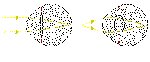
Light from a single point of a distant object and light from a single point of a near object being brought to a focus.
Other articles regarding eye anatomyAnnulus of Zinn, Conjunctiva, Macula, Nictitating membrane, Schlemm's canal, Trabecular meshwork.
Cytology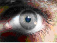
This image clearly shows the pupil, iris, and blood vessels of the human eye.
The retina contains two forms of photosensitive cells important to vision — rods and cones. Though structurally and metabolically similar, their function is quite different. Rod cells are highly sensitive to light allowing them to respond in dim light and dark conditions. These are the cells which allow humans and other animals to see by moonlight, or with very little available light (as in a dark room). However, they do not distinguish between colours.
This is why the darker conditions become, the less colour objects seem to have. Cone cells, conversely, need high light intensities to respond and have high visual acuity. Different cone cells respond to different wavelengths of light, which allows an organism to see colour.
The differences are useful; apart from enabling sight in both dim and light conditions, humans have given them further application. The fovea, directly behind the lens, consists of mostly densely-packed cone cells. This gives humans a highly detailed central vision, allowing reading, bird watching, or any other task which primarily requires looking at things. Its requirement for high intensity light does cause problems for astronomers, as they cannot see dim stars, or other objects, using central vision because the light from these is not enough to stimulate cone cells. Because cone cells are all that exist directly in the fovea, astronomers have to look at stars through the "corner of their eyes" (averted vision) where rods also exist, and where the light is sufficient to stimulate cells, allowing the individual to observe distant stars.
Rods and cones are both photosensitive, but respond differently to different frequencies of light. They both contain different pigmented photoreceptor proteins. Rod cells contain the protein rhodopsin and cone cells contain different proteins for each colour-range. The process through which these proteins go is quite similar — upon being subjected to electromagnetic radiation of a particular wavelength and intensity (ie. a colour visible light), the protein breaks down into two constituent products. Rhodopsin, of rods, breaks down into opsin and retinal; iodopsin of cones breaks down into photopsin and retinal. The opsin in both opens ion channels on the cell membrane which leads to the generation of an action potential (an impulse which will eventually get to the visual cortex in the brain).
This is the reason why cones and rods enable organisms to see in dark and light conditions — each of the photoreceptor proteins requires a different light intensity to break down into the constituent products. Further, synaptic convergence means that several rod cells are connected to a single bipolar cell, which then connects to a single ganglion cell and information is relayed to the visual cortex. Whereas, a single cone cell is connected to a single bipolar cell. Thus, action potentials from rods share neurons, where those from cones are given their own. This results in the high visual acuity, or the high ability to distinguish between detail, of cone cells and not rods. If a ray of light were to reach just one rod cell this may not be enough to stimulate an action potential. Because several "converge" onto a bipolar cell, enough transmitter molecules reach the synapse of the bipolar cell to attain the threshold level to generate an action potential.
Furthermore, color is distinguishable when breaking down the iodopsin of cone cells because there are three forms of this protein. One form is broken down by the particular EM wavelength that is red light, another green light, and lastly blue light. In simple terms, this allows human beings to see red, green and blue light. If all three forms of cones are stimulated equally, then white is seen. If none are stimulated, black is seen. Most of the time however, the three forms are stimulated to different extents — resulting in different colours being seen. If, for example, the red and green cones are stimulated to the same extent, and no blue cones are stimulated, yellow is seen. For this reason red, green and blue are called primary colours and the colours obtained by mixing two of them, secondary colors. The secondary colours can be further complimented with primary colours to see tertiary colors.
Acuity

Closeup of a hawk's eye.
Visual acuity can be measured with several different metrics.
Cycles per degree (CPD) measures how much an eye can differentiate one object from another in terms of degree angles. It is essentially no different from angular resolution. To measure CPD, first draw a series of black and white lines of equal width on a grid (similar to a bar code). Next, place the observer at a distance such that the sides of the grid appear one degree apart. If the grid is 1 meter away, then the grid should be about 8.7 millimeters wide. Finally, increase the number of lines and decrease the width of each line until the grid appears as a solid grey block. In one degree, a human would not be able to distinguish more than about 12 lines without the lines blurring together. So a human can resolve distances of about 0.93 millimeters at a distance of one meter. A horse can resolve about 17 CPD (0.66 mm at 1 m) and a rat can resolve about 1 CPD (8.7 mm at 1 m).
A diopter is the unit of measure of optical power.
------------------------------------------------------------------------------------------------
Want fun then why wait? Click on any one of URL
http://blog4people.coz.in/ http://jokes4children.coz.in/ http://adsense.coz.in/ http://kannada.coz.in/ http://ourmysore.coz.in/ http://wowbangalore.coz.in/ http://wowchennai.coz.in/ http://bhshistory.coz.in/ http://karaga.coz.in/


